Myocardial Architecture
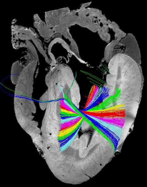
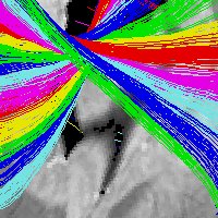
Fibertracking in giraffe heart using DTI MRI
Myocardial Architecture
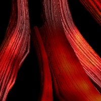
Cardiac anatomy is the foundation of both normal and abnormal cardiac function. The intricate microstructure of the heart and its relationship with cardiac function has been debated for decades, without current consensus. Although this appears to be fundamental information, astonishingly many clinical procedures, interpretations, diagnoses and research studies are based on poor anatomical knowledge, stemming from inaccurate textbook descriptions and anatomically unfounded ideologies. There are still inconsistencies with regards to the nomenclature of cardiac structure, this matter needs to be resolved; it is important that clinicians around the world speak the same anatomical language. To improve diagnosis and to devise rational treatments heart disease, we first need to understand the micro-anatomical changes that underlie normal and abnormal contraction and relaxation across the heart.
Utilising the technique of diffusion tensor imaging we investigate the micro-anatomy of both the healthy and the diseased myocardium. Diffusion tensor imaging is a subtype of magnetic resonance assessing the spontaneous diffusion of water molecules in tissues. It has been shown that the diffusion of water follows the orientation of the cardiomyocytes, thus we assess the diffusion direction as a surrogate measure of myocyte orientation.
We have described the anatomy of both normal and diseased hearts mainly in porcine and ovine animal models of congenital heart disease. Furthermore, we have undertaken a quest to elucidate the differences in myocardial architecture between mammals. This is done in a search for alternative ideas applicable when surgically correcting congenital heart disease. This research has lead us to the investigation of many different species of which giraffes are of high interest due to their unique blood pressure regulation capabilities.
PROJECTS
Myocardial Architecture
Fiber tractography of peripheral nervous tissue in a forensic setting
Telemetric nerve recordings from peripheral nerves