Non-invasive tracking of cells
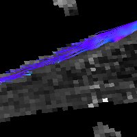
Tracking of SPIO-labelled axolotl blastema cells using non-invasive MRI
Non-invasive tracking of cells
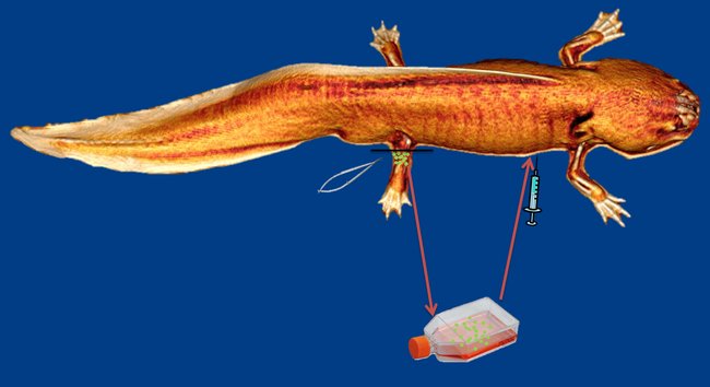
Before regenerative therapies can ever come to play, an appropriate monitoring technology has to be developed that ensures non-invasive follow up examinations of patients undergoing therapy. In studies evaluating the effectiveness of stem cell-based regenerative therapies, monitoring has traditionally relied on histological techniques. In order to detect the presence of cells within the region of interest, research animals are usually sacrificed and subjected to biopsy and histology. Even though these methods and techniques are valuable in a research setting, they preclude non-invasive in vivo assessment and longitudinal tracking of therapeutic progress.
Stem or progenitor cell fate can be monitored using an alternative method by labelling cells of interest with non-toxic super-paramagnetic iron oxide particles (SPIOs) that allow for in vivo cell tracking using magnetic resonance imaging (MRI). On the assumption that SPIOs are in fact non-toxic to the labelled cells, this methodology is minimally invasive and completely safe due to the harmless nature of MRI. SPIOs are either internalised by the endosomal-lysosomal pathway or bind to the surface of cells, and due to their magnetic properties, they increase the magnetic susceptibility and decrease the MRI-measured properties of water, the spin-spin (T2) and to some degree the spin-lattice (T1) relaxation times. Labelled SPIOs have successfully been used to track stem cell migration and quantify the amount of cells arriving in the target zone. At present the SPIO labelling technique has been applied in a number of preclinical studies, but to never in a system with true intrinsic regenerative capacity.
The purpose of this study is to introduce SPIO labelling for cell tracking in a truly regenerative environment, the regenerating limb of the axolotl, and subsequently investigate the homing effect of blastema cells to the regenerative zone when applied intravascularly.
PROJECTS
Cardiac regeneration
Non-invasive tracking of cells
Spinal cord regeneration – a contusion trauma model