MRI, ultrasound and bubbles
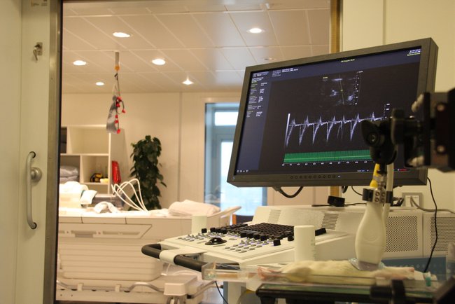
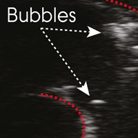
Changes in basic MRI parameters (T1, T2 and T2*) in the rat brain were correlated to bubble formation in the pulmonary artery assessed using ultrasonography. The imaging compatible pressure chamber is visible in the background.
MRI, ultrasound and bubbles
The development of intra-vascular and tissue bubbles has long been considered the pivotal pathological event in the development of adverse symptoms of decompression sickness (DCS) after diving. While it is generally accepted that absence of bubbles and low bubble grades indicate a low risk of developing DCS, both in humans and animals, the relationship between the occurrence of bubbles and development of DCS-symptoms is complex. It is acknowledged that the risk for DCS development is statistically elevated the higher the number of precordial bubbles observed, but nonetheless, high number of bubbles are found in DSC-asymptomatic human divers.
The brain and central nervous system are often affected in more serious cases of DCS and bubbles have been reported to cause stroke-like symptoms and to resemble mild traumatic brain injury. Symptoms as dizziness, headaches, memory changes, poor concentration, coordination deficiencies, numbness, and paraesthesia (i.e. a pricking, burning, tingling or numbing feeling of extremities) are among the symptoms often reported in recreational diving accidents involving CNS symptoms.
In this study the developed imaging-compatible pressure chamber system was used to characterise changes in basic MRI-parameters (T1, T2 and T2*) in the rat brain during pressurisation and decompression. These changes were correlated the development of bubbles in the venous vasculature visualised through echo graphic brightness-mode images using micro-ultrasound imaging.
Overall, the study concluded that MRI relaxometry is a sensitive method for registering cerebral effects following pressurisation and decompression in rats.
Projects
A CT, PET and MRI compatible pressure chamber for baromedical research
MRI, ultrasound and bubbles
Looking into treatment of decompression sickness