Fiber tractography of peripheral nervous tissue in a forensic setting
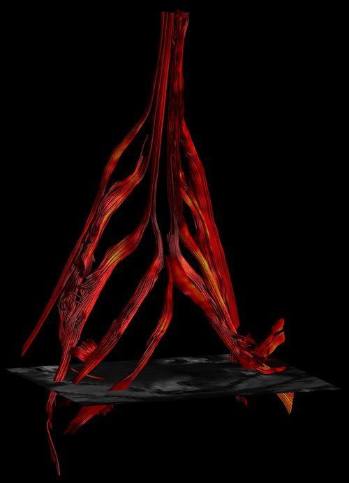
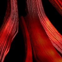
The lumbosacral (L3-S2) nerves in a post mortem subject. The colour encoding is according to the fractional anisotropy
Fiber tractography of peripheral nervous tissue in a forensic setting
Nowadays imaging modalities are used often in a forensic setting to visualise the body non-invasively and obtain additional information which can be helpful in identifying cause of death. Magnetic resonance imaging (MRI) can be used in this area to investigate soft tissue and organ injuries.
The potential value in forensic sciences of more advanced techniques such as diffusion tensor imaging (DTI) remains unexplored. Peripheral nerves are hard to dissect during regular autopsy, and this is therefore a rather neglected area. DTI has the potential to identify nervous structures with great detail and can help in providing morphological information using fiber tractography.
It can also provide additional information consisting of different diffusion parameters to characterize nervous tissue properties. Spinal or neck traumatic injuries which stem from a sudden, traumatic blow that fractures one or more of the vertebrae and surrounding nervous tissue can be hard to identify during conventional autopsy.
This techniques might be helpful in these trauma cases but could also be beneficial in for example homicides. However, its application in a post mortem setting can be challenging since there are different degenerative processes starting soon after death. In this research we explore the potential of DTI to be used as a tool to assess peripheral nervous tissue in non-fixed post mortem cases.
PROJECTS
Myocardial Architecture
Fiber tractography of peripheral nervous tissue in a forensic setting
Telemetric nerve recordings from peripheral nerves